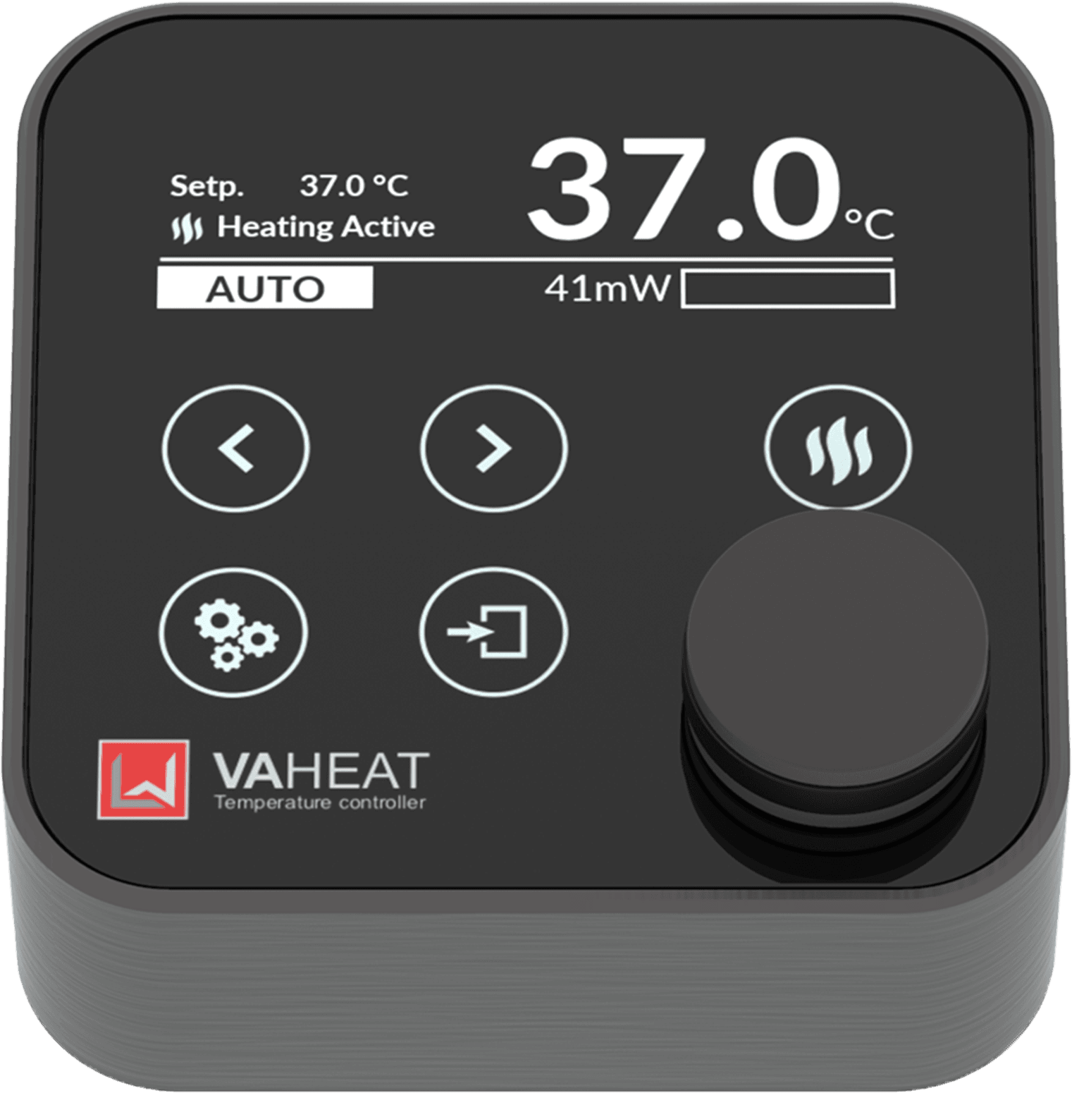
Dynamic Temperature Control
Microscopic temperature control of your sample volume for high-resolution optical microscopy. Just set the temperature and start your measurement. Let VAHEAT do the rest.
about temperature control in high resolution microscopy.
Key Features
VAHEAT is a precise temperature control unit for optical microscopes. It combines local heating with direct temperature sensing in the sample volume. This allows for fast and precise temperature adjustment with heating rates up to 100°C/s while maintaining highest temperature precision. Made for investigations of temperature-sensitive processes in life sciences and material research.
Extreme temperature stability on long (hours to days) and short (seconds to minutes) time scales down to 0.01°C (rms). External temperature variations due to air flow, fluid exchange etc. will be detected and compensated via direct temperature feedback inside the sample volume.
Extend your experimental temperature range to 100°C (standard range) or even up to 200°C (extended range) depending on your experimental needs. The standard range version is compatible with oil-immersion systems while the extended range version can be operated with air objectives.
No optical aberration up to 80 °C with the highest numerical aperture objectives on the market. Perfectly suited for single molecule and super resolution studies using state-of- the-art methods (STORM, STED, TIRF, etc.). For more see Technical Performance.
VAHEAT lets you control the temperature inside the field of view independently from the type of microscope objective or the objective’s temperature. The system is designed as standalone unit without the need for any additional modifications to the optical setup (e.g objective heater) in order to avoid a temperature sink in your field of view. Additionally, the specific design of our smart substrates ensures that the objective’s performance is not altered even at higher temperatures.
VAHEAT is equipped with four heating modes for different experiments depending on your needs. Modes for fast heating, auto-compensated heating, or well-defined temperature profiles are available.
No thermal drifts or vibrations even at elevated temperature allow precise single molecule localization. We designed VAHEAT to be compatible with all commercial microscopes. No further modifications of your setup are needed. Its fast thermal response allows for nearly instantaneous thermalization tremendously reducing the waiting times as usual with conventional heating systems.
Components
Control Unit
- STANDARD RANGE
- EXTENDED RANGE
Heating Power
<2500 mW
Max. temperature
105 °C.
Smart Substrates
SmS and SmS-R
Applications
High and super resolutions studies
The extended range version is meant for studies up to 200 °C. The maximal heating power of 5000 mW boosts your sample temperature to your desired setpoint even when larger thermal loads are attached. This system is made for investigations using air
spaced microscope objective for studying e.g. phase transitions or diffusional behavior.
Heating Power
<5000 mW
Max. temperature
200 °C
Smart Substrates
SmS, SmS-R and SmS-E
Applications
High resolutions studies
VAHEAT can be operated in four different modes that will ideally fit your experimental needs.
- AUTO
- DIRECT
- SHOCK
- PROFILE
The SHOCK mode resembles a timed DIRECT mode. For a defined period, a certain heating power will be applied to the sample volume without any feedback loop being active. This allows to exploit the fast heating dynamics of VAHEAT to send heat shocks into your sample.
Smart Substrates
Dimension: 18x18x0.17 (± 0.05) mm³
Temp. range: RT - 105°C
The standard range smart substrates are optimized for high-resolution studies similar to #1.5 coverslips. They have a size of 18 x 18 mm² and a thickness of 170 µm. We specify their full functionality up to 100 °C with heating powers up to 2.5 W. The flat substrates are ideally suited to integrate a reservoir, study thin films or use an own design of a microfluidic chamber.
Dimension: 18x18x0.17 (± 0.05) mm³
Temp. range: RT - 105°C
Volume: 100 - 600 µL
The smart substrates equipped with a reservoir made of silicone are meant for investigating liquid samples of a maximal volume of 600 µL. All materials used are non-cytotoxic and meet the highest standards for live-cell imaging. The SmS-R are suited for micro stage top incubation. Upon request we can provide additional glass slides for sealing the reservoir.
Dimension: 18x18x0.5 mm³
Temp. range: RT - 200°C
The extended range smart substrates are made for temperatures up to 200 °C. They are 500 µm thick, similar to #5 coverslips and are compatible with the extended range control unit.
Dimension: 18x18x0.17 (± 0.05) mm³
Temp. range: RT - 105°C
The standard range smart substrates are optimized for high-resolution studies similar to #1.5 coverslips. They have a size of 18 x 18 mm² and a thickness of 170 µm. We specify their full functionality up to 100 °C with heating powers up to 2.5 W. The flat substrates are ideally suited to integrate a reservoir, study thin films or use an own design of a microfluidic chamber.
Dimension: 18x18x0.17 (± 0.05) mm³
Temp. range: RT - 105°C
Volume: 100 - 600 µL
TThe smart substrates equipped with a reservoir made of silicone are meant for investigating liquid samples of a maximal volume of 600 µL. All materials used are non-cytotoxic and meet the highest standards for live-cell imaging. The SmS-R are suited for micro stage top incubation. Upon request we can provide additional glass slides for sealing the reservoir.
Dimension: 18x18x0.5 mm³
Temp. range: RT - 200°C
The extended range smart substrates are made for temperatures up to 200 °C. They are 500 µm thick, similar to #5 coverslips and are compatible with the extended range control unit.
Microscope Adapter
The heart of the VAHEAT system are the smart substrates that integrate a transparent thin film heating element with a highly sensitive temperature probe. The smart substrates with a heated area of 5 x 5 mm2 are inserted into the substrate holder that is connected to the control unit. As soon as the connector head establishes the connection between smart substrate and control unit, the temperature is being measured and you can start heating.
The microscope adapter fits into most of the common microscope stages due to its size of 75 mm x 25 mm (3” x 1”). Its slim design with a maximal height of 11 mm allows unrestricted access from above. The microscope adapter is thermally insulated from the heated area and stays at room temperature even for sample temperatures of 200 °C. Simply prepare your sample on the smart substrates, insert it into the microscope adapter and position it onto your translation stage to start your measurement. VAHEAT takes care of precise temperature control.
The User Interface
The platform independent user interface (UI) allows to remotely control the VAHEAT device, program arbitrary temperature profiles and stream temperature data to the local hard disk. You can use it for precise and live control of sample temperature and current heating power. Simply connect the control unit via USB to the computer and start the measurement remotely.
This is what our customers think about VAHEAT
“One part of my laboratory works on transcription factors in the germline of C.elegans and liquid-liquid phase separation. Temperature dependence is one of the better ways to show whether protein foci are formed by a phase transition mechanism or not. I have done temperature dependent experiments in the past with a home built system and I know how difficult it is. Compared to that, the VAHEAT system is really easy to use across many microscopes and samples. We use it for C.elegans, zebrafish and single cells."
Dr. Senthil Arumugam
EMBL Australia/Monash University
“I had a chance to work with VAHEAT at the Woods Hole Physiology course. We combined VAHEAT with our custom microfluidic devices and imaged live archaeal cells for single-cell growth curves of many different species. We leveraged VAHEAT fine temperature controls to create gradient ramps over long periods of time time to optimize the growth of mixed populations. VAHEAT is a great tool for cell biologists that rely on challenging temperature ranges, and it can be even better with a larger heated surface area to allow multi-fluidic channels for high-throughput imaging."
Dr. Alexandre Bisson
Brandeis University
"VAHEAT allows precise and fast temperature control in our TIRF measurements where we are investigating switchable DNA origami mechanisms.“
Prof. Hendrik Dietz
TU Munich
“At the beginning I was a bit sceptical about the substrates. However, they can be cleaned and reused. We have tested several heating systems that can heat above 37°C. This one was by far our favourite."
Dr. Kerstin Göpfrich
MPI for Medical Research, Heidelberg
"We use the VAHEAT to bring low molecular weight polymers to temperatures just above their glass transition temperature to study molecular motions and dynamic heterogeneity at the single molecule level in these systems. The fact that the VAHEAT allows us to achieve and maintain the temperature control needed on a coverslip in turn allows us to simultaneously perform imaging with the highest numerical aperture objectives available, maximizing collection of photons and limiting localization error of fluorescent probes. This ability facilitates characterization of translational mobility in these complex systems."
Prof. Laura Kaufman
Columbia University, New York City
“The instructions and equipment were very clear and intuitive to use. We very much enjoyed the magnets that lock components into place. In use, the temperature changed very rapidly according to the VAHEAT reading, though I allowed some extra time to equilibrate as my material is not very conductive. I need to carry out further analysis, but I am seeing what I expected to see in my results. The biggest advantage was indeed that I didn’t see any thermal drift: I didn’t need to refocus during my experiments, which is fantastic because I wanted to see fluorescence emission intensity in a single controlled plane. Overall, I think very highly of the VAHEAT and it was immensely helpful to me."
Dr. Liz Birchall
University of Nottingham
“Thanks to VAHEAT, we were able to perform live imaging of cell divisions in thermophilic Archaea for the first time in our lab. Another application, where we successfully used the instrument was to capture the swimming behaviour of Sulfolobus."
Dr. Marleen van Wolferen
University of Freiburg
“I tested VAHEAT in my experiments, where I image fluorescent thermoresponsive polymers. I found the device very useful and decided to purchase it. I combined the Smart Substrates with silicon spacers and another coverslip to form a custom small volume imaging chamber ideally suited for my samples."
Serena Teora
Radboud University
“I have tested VAHEAT in live cell imaging and for in vitro experiments with protein condensation. The device performance convinced me and I think that it could be another flexible option for temperature control for our users. I liked VAHEAT also as a “mere” temperature sensor. Perfect for quality control options for facility people, but also for individual users."
Dr. Josef Gotzmann
Head of BioOptics Facility, Max Perutz labs
“We were testing VAHEAT for two photon imaging of cellular processes in neurons. We were able to grow neuronal cultures on the Smart Substrates, although only mixed cultures and not pure primary neurons. The device itself worked well. I recorded SHG images and temperature was stable during the recordings. Personally I find this a quite handy device."
Tobias Martens
Cell Imaging Core at KU Leuven
“We are running experiments on DNA-modified self-assembling colloidal particles. We operate in a very narrow temperature range of 0.5°C around the melting temperature of our DNA oligonucleotides. For this type of measurement we have found VAHEAT heating stage very useful. In our setup, we used a bare VAHEAT substrate and glued a capillary on top of it. We filled the capillary with our solution, which allowed us to use a small sample volume, maintain very stable temperature over long periods of time, not having to worry about temperature drift or evaporation. This setup gave us sufficient control over the temperature to manipulate crystallization in our colloidal system."
Dr. Bas van Ravensteijn
Eindhoven University of Technology (Currently: Utrecht University)
“We tested VAHEAT in live cell imaging to heat shock our yeast strains containing temperature sensitive alleles and it helped our meiosis research. I have done many temperature shift experiments with using different systems and it can be quite challenging. Personally, I find the VAHEAT system is really easy to use compared to that. It can be even better with a larger and divided surface area to allow imaging of different strains at the same time."
Dr. Tugce Oz Yoldas
MPI of Biochemistry, Martinsried
“Thank you for providing us with such a reliable way to control the temperature of our samples. You revolutionized the way we do heat shock!"
Dr. Linda Rubio
LSU Health, Shreveport
This is what our customers think about VAHEAT
“One part of my laboratory works on transcription factors in the germline of C.elegans and liquid-liquid phase separation. Temperature dependence is one of the better ways to show whether protein foci are formed by a phase transition mechanism or not. I have done temperature dependent experiments in the past with a home built system and I know how difficult it is. Compared to that, the VAHEAT system is really easy to use across many microscopes and samples. We use it for C.elegans, zebrafish and single cells."
Dr. Senthil Arumugam
EMBL Australia/Monash University
“I had a chance to work with VAHEAT at the Woods Hole Physiology course. We combined VAHEAT with our custom microfluidic devices and imaged live archaeal cells for single-cell growth curves of many different species. We leveraged VAHEAT fine temperature controls to create gradient ramps over long periods of time time to optimize the growth of mixed populations. VAHEAT is a great tool for cell biologists that rely on challenging temperature ranges, and it can be even better with a larger heated surface area to allow multi-fluidic channels for high-throughput imaging."
Dr. Alexandre Bisson
Brandeis University
"VAHEAT allows precise and fast temperature control in our TIRF measurements where we are investigating switchable DNA origami mechanisms.“
Prof. Hendrik Dietz
TU Munich
“At the beginning I was a bit sceptical about the substrates. However, they can be cleaned and reused. We have tested several heating systems that can heat above 37°C. This one was by far our favourite."
Dr. Kerstin Göpfrich
MPI for Medical Research, Heidelberg
"We use the VAHEAT to bring low molecular weight polymers to temperatures just above their glass transition temperature to study molecular motions and dynamic heterogeneity at the single molecule level in these systems. The fact that the VAHEAT allows us to achieve and maintain the temperature control needed on a coverslip in turn allows us to simultaneously perform imaging with the highest numerical aperture objectives available, maximizing collection of photons and limiting localization error of fluorescent probes. This ability facilitates characterization of translational mobility in these complex systems."
Prof. Laura Kaufman
Columbia University, New York City
“The instructions and equipment were very clear and intuitive to use. We very much enjoyed the magnets that lock components into place. In use, the temperature changed very rapidly according to the VAHEAT reading, though I allowed some extra time to equilibrate as my material is not very conductive. I need to carry out further analysis, but I am seeing what I expected to see in my results. The biggest advantage was indeed that I didn’t see any thermal drift: I didn’t need to refocus during my experiments, which is fantastic because I wanted to see fluorescence emission intensity in a single controlled plane. Overall, I think very highly of the VAHEAT and it was immensely helpful to me."
Dr. Liz Birchall
University of Nottingham
“Thanks to VAHEAT, we were able to perform live imaging of cell divisions in thermophilic Archaea for the first time in our lab. Another application, where we successfully used the instrument was to capture the swimming behaviour of Sulfolobus."
Dr. Marleen van Wolferen
University of Freiburg
“I tested VAHEAT in my experiments, where I image fluorescent thermoresponsive polymers. I found the device very useful and decided to purchase it. I combined the Smart Substrates with silicon spacers and another coverslip to form a custom small volume imaging chamber ideally suited for my samples."
Serena Teora
Radboud University
“We were testing VAHEAT for two photon imaging of cellular processes in neurons. We were able to grow neuronal cultures on the Smart Substrates, although only mixed cultures and not pure primary neurons. The device itself worked well. I recorded SHG images and temperature was stable during the recordings. Personally I find this a quite handy device."
Tobias Martens
Cell Imaging Core at KU Leuven
“I have tested VAHEAT in live cell imaging and for in vitro experiments with protein condensation. The device performance convinced me and I think that it could be another flexible option for temperature control for our users. I liked VAHEAT also as a “mere” temperature sensor. Perfect for quality control options for facility people, but also for individual users."
Dr. Josef Gotzmann
Head of BioOptics Facility, Max Perutz labs
“We tested VAHEAT in live cell imaging to heat shock our yeast strains containing temperature sensitive alleles and it helped our meiosis research. I have done many temperature shift experiments with using different systems and it can be quite challenging. Personally, I find the VAHEAT system is really easy to use compared to that. It can be even better with a larger and divided surface area to allow imaging of different strains at the same time."
Dr. Tugce Oz Yoldas
MPI of Biochemistry, Martinsried
"Thank you for providing us with such a reliable way to control the temperature of our samples. You have revolutionized the way we do heat shock!"
Dr. Linda Rubio
LSU Health, Shreveport
Technical Performance
- IMAGING QUALITY
- TEMPERATURE STABILITY
- HEATING RATES
100x, NA=1.46, oil immersion objective
40x, NA=0.4, air objective
Temperature fluctuations over time
Temperature step response
Compatible Imaging Techniques
Total internal reflection microscopy (TIRM)
Confocal microscopy
Interferometric scattering microscopy (iSCAT)
Atomic force microscopy (AFM)
Super resolution methods (SIM, STORM, PALM, PAINT, STED)
Widefield microscopy
Total internal reflection microscopy (TIRM)
Confocal microscopy
Interferometric scattering microscopy (iSCAT)
Atomic force microscopy (AFM)
Super resolution methods (SIM, STORM, PALM, PAINT, STED)
Widefield microscopy
Application Examples
- LIVE CELL IMAGING
- DNA
- MICROFLUIDICS
- PHASE TRANSITIONS
- NEUROSCIENCE
- AFM
Living organisms are highly sensitive to changes in their environmental conditions, especially to temperature. VAHEAT ensures reliable and precise temperature control while transfer and during imaging. Investigating temperature sensitive processes of cellular behavior, such as Ca2+ activity in multicellular tumor spheroids or thermal stimulation of neurons has never been this easy.
Exploit VAHEAT ‘s capabilities as micro stage top incubator.
CAtegories:
Cell Biology | Neuroscience | Medicine | Stage Top Incubator
CAtegories:
CAtegories:
Nanotechnology | Biochemistry | Biotechnology
CAtegories:
CAtegories:
CAtegories:
Selected publications
Live imaging of thermophilic bacteria under confinement
C. Molinaro et al., RSC Advances (2021)
Guillaume Baffou’s lab from Institut Fresnel used VAHEAT to image growing thermophilic bacteria at 60°C and 70°C under spatial confinement. They found out that what is good for E. coli does not necessarily satisfy other non-model organisms. Most aerobic bacteria need for successful growth more oxygen than is available in confined geometries.
Live imaging of meiosis in yeast
V. Mengoli et al., EMBO journal (2021)
Wolfgang Zachariae‘s lab from the Max Planck Institute for Biochemistry used temperature-sensitive alleles in yeast to study chromosome separation during meiosis. Shifting yeast from 25°C to 37°C while simultaneously doing live cell imaging? Ideal application for our instrument!
Single-molecule TIRF measurements of DNA origami
P. Stömmer et al., Nature Communications (2021)
The laboratory of Hendrik Dietz from Technical University Munich built a macromolecular transport system out of DNA origami. VAHEAT was used for precise temperature control in the single-molecule TIRF measurements. The article was published in Nature Communications.
iSCAT microscopy of nanoparticles
A. Kashkanova et al., Nature Methods (2021)
The laboratory of Vahid Sandoghdar at the Max Planck Institute for the Science of Light keeps pushing the limits of iSCAT microscopy.
In their new preprint they used VAHEAT to characterise temperature dependence of diffusion constant of gold nanoparticles.
Videos
Temperature sensitive microscopy in life sciences
Watch our webinar from November 16th 2022 focused on different uses of VAHEAT in cell biology, membrane biophysics and chemistry. Dr. Arthur Charles-Orszag presented a study of archaeal cell motility. Prof. Sarah Veatch talked about exciting data on cross talk between membrane phase transition and protein LLPS. Serena Teora showed her results with thermo-responsive micromotors.
VAHEAT as a precise temperature reference
Watch our webinar from March 23rd 2022 focused on using VAHEAT as a temperature calibration tool. Andrew York (Calico), Guillaume Baffou (Institut Fresnel) and Pascal Lorentz (University of Basel) presented very different projects, where VAHEAT’s precision made a difference and allowed them to make sense out of their imaging data or to discover imprecise temperature control in their light microscopy facility.
Imaging colloids - focus on temperature
Watch our webinar from November 10th 2021 focused on colloids and VAHEAT. Ahu Gümrah Dumanli-Parry (University of Manchester) talked about anisotropic self-assembly to produce optically chiral active matter and structurally coloured materials. Bas van Ravensteijn (Utrecht University) presented his research on synthesis of colloidal systems through DNA mediated self-assembly.
Live cell imaging - focus on temperature
Watch our webinar from 13th October 2021 focused on Live cell imaging and VAHEAT. Senthil Arumugam (Monash University) presented his research of endosomal maturation in human cells as imaged by lattice light sheet microscopy. Alexandre Bisson (Brandeis University, USA) talked about challenges in capturing the cell biology of Archaea live in a microscope.
VAHEAT as stage top incubator for live cell imaging
Long-term live cell imaging of normal human dermal fibroblasts (NDHF) that are stained with SIR-tubulin. The cells are incubated using only a Smart Substrate with Reservoir (SmS-R) and are continuously imaged for more than 18 hrs. No need for large incubation chambers, objectives heater or prior setup calibration!
Heat shock of live cells
Human dermal fibroblasts (NDHF) that are stained with SIR-tubulin are held at 37 °C using VAHEAT as a stage top incubator. The sample volume is continuously imaged while sending heat shocks of 45°C for 30 s and 55°C for 30 s into the sample that result in cell death. The imaging quality is maintained during and the sample position is not shifted.
Temperature control with VAHEAT
Inverted microscope with oil immersion objective is equipped with a VAHEAT system. Only the smart substrate is temperature controlled (no objective heater). The infrared camera shows a homogeneous temperature distribution inside the heated area although the objective is in thermal contact with the smart substrate.
FAQs
Content
The standard range version can issue heating powers up to 2.5 W and is working with smart substrates of 170 μm thickness (#1.5H) as required for high NA immersion microscope objectives. The high-temperature version can be operated with smart substrates of 500 μm thickness (#5) while allowing heating powers of up to 5.0 W.
Yes! VAHEAT is compatible with vacuum chambers. You might require a specific cable feedthrough but there will be no outgassing when operating VAHEAT inside a vacuum chamber. Contact us if you need more information.
Yes! We specifically designed VAHEAT
for high- and super-resolution microscopy. The smart substrates have a thickness of 170 μm and are thus compatible with almost any commercially available high Numerical Aperture (NA) objective needed for super- resolution microscopy.
The conventional thermal problems that arise in super-resolution microscopy when working at elevated temperature are fully avoided when working with VAHEAT. We also measured the optical performance with a high NA objective as function of temperature. You can check out the data here.
You cannot believe this? Get a demo system and test it yourself!
From internal measurements we can conclude that the sample heating has no effect on the image quality and therefore on the point spread function (PSF) for temperatures between room temperature and 100 °C.
We recommend oil and water based immersion medium. Please avoid glycerine as immersion medium as it will attack the smart substrates and depreciate their functionality.
Yes! We offer a five-week test period. Reach out to us!